[TargetMol Star Molecule] - TD139: A pivotal probe targeting Galectin-3 for advancing pulmonary fibrosis and immune modulation research
■ 1. Background
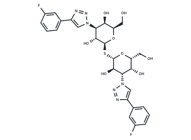
▲ TD139 (T5121),CAS: 1450824-22-2
TD139 (T5121), CAS: 1450824-22-2 is a small-molecule inhibitor of galectin-3 (Gal-3) developed for inhaled delivery to the airways, widely used as a chemical probe to interrogate Gal-3–dependent fibrotic mechanisms in the lung, including models relevant to idiopathic pulmonary fibrosis. Gal-3 is a β-galactoside–binding lectin that operates at the nexus of the galectin pathway, functioning both extracellularly—by cross-linking glycosylated receptors and matrix ligands to assemble signaling lattices—and intracellularly as a scaffolding factor that modulates cell survival and transcriptional programs [1]. In the fibrotic lung, Gal-3 facilitates profibrotic signaling, notably by potentiating transforming growth factor-β (TGF-β) activity and sustaining macrophage–fibroblast crosstalk that drives myofibroblast differentiation and extracellular matrix (ECM) deposition [2].
Mechanistically, TD139 targets the carbohydrate recognition domain of Gal-3, competitively blocking its binding to β-galactoside–containing glycoconjugates on cell-surface receptors and ECM components. By interrupting Gal-3–mediated lattice formation and receptor clustering, TD139 dampens stabilization and amplification of signaling nodes that rely on glycan-dependent organization, including TGF-β receptor complexes and other glycoprotein receptors implicated in fibroblast activation [1,2]. This results in reduced propagation of canonical profibrotic signaling (for example, attenuation of TGF-β/SMAD transcriptional drive) and downstream expression of genes encoding contractile and matrix proteins. Because Gal-3 also modulates macrophage phenotype and secretory profiles, TD139 can shift the cytokine milieu that conditions fibroblast behavior, thereby influencing ECM remodeling trajectories in fibrotic microenvironments [2].
Within the broader galectin pathway, key components include Gal-3 itself, multivalent glycoconjugate ligands presented on receptors and the ECM, and the dynamic lattices that control receptor residency, endocytosis, and signal duration. TD139 functionally disassembles this architecture by preventing Gal-3 from bridging glycan moieties, decreasing receptor nanocluster stability and curtailing signal persistence. In pulmonary research, inhaled administration is advantageous, enabling high local target engagement in airway and alveolar compartments while allowing pharmacodynamic readouts such as changes in bronchoalveolar lavage Gal-3 activity, fibroblast activation markers, and collagen content in tissue explants.
TD139 is therefore leveraged as a pathway-selective tool to: (i) delineate Gal-3’s contribution to TGF-β–centric profibrotic circuits; (ii) parse glycan-dependent receptor organization effects on signal transduction; (iii) interrogate macrophage–fibroblast communication in fibrotic niches; and (iv) benchmark Gal-3 target engagement alongside transcriptional and histologic endpoints. In vitro, it is used in primary human lung fibroblasts, epithelial cells, and macrophages to quantify shifts in activation markers and ECM gene programs. In vivo and ex vivo lung systems, including precision-cut tissue slices, employ TD139 to map dose–response relationships between Gal-3 inhibition and fibrosis-relevant biomarkers. Collectively, these applications position TD139 as a focused modulator of the galectin pathway for probing the mechanistic underpinnings of fibrogenesis in the respiratory tract [1,2].
■ 2. Literature review
■ 2.1 Galectin-3 mediates lysosome-related inflammation within monocyte-derived macrophages in a mouse model of ischemic brain injury
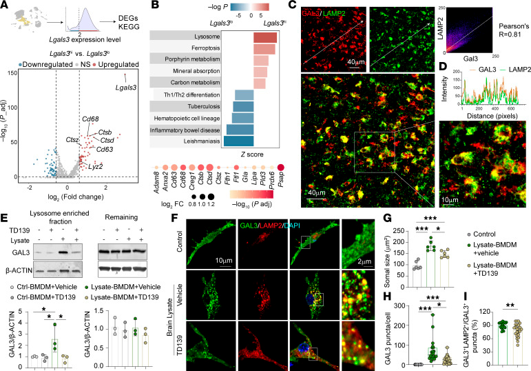
▲ (A) Experimental scheme illustrating the analysis of differentially expressed genes (DEGs) between Lgals3^hi and Lgals3^lo MDMø 3 d after tMCAO. MDMø were divided into 2 groups based on Lgals3 expression level >2 or ≤2. DEGs were identified by comparing the 2 groups. A volcano plot depicts the DEGs [log (fold change) >0.59 or < –0.59, Bonferroni adjusted P value <0.05] between Lgals3^hi and Lgals3^lo MDMø. (B) KEGG enrichment analysis was performed on the DEGs using Metascape, showing the 10 KEGG pathways predicted to be upregulated or downregulated (z score ≥5 or z score ≤5). Corresponding dot plot shows upregulated genes involved in the lysosome term in the Lgals3^hi group. (C) Representative LAMP2 and GAL3 staining in ischemic brains 3 d after tMCAO. Right panel shows Pearson's correlation analysis of GAL3 and LAMP2 signals. Scale bars: 40 μm, 10 μm (zoom). (D) Fluorescence intensity profiles of GAL3 (red) and LAMP2 (green) signals for objects crossing the white arrows in C. (E) Immunoblot and quantitative analysis of GAL3 expression in lysosome-enriched fraction and the remaining cell lysates (without lysosome) from cultured BMDMs under the indicated conditions. N = 3/condition. (F) Representative images of GAL3 and LAMP2 staining in control and brain lysate-treated BMDMs with or without TD139 (10 μM). (G) The average somal size of BMDMs. N = 6 per condition. (H) The numbers of GAL3+ puncta per cell. (I) The percentages of GAL3+LAMP2+ puncta among total GAL3+ puncta under the indicated conditions. N = 22–23 randomly selected cells per condition from 3 independent experiments. *P < 0.05, **P < 0.01; ***P < 0.001. Two-tailed, unpaired Student's t test (I), 1-way ANOVA and Bonferroni's test (E and G), or Kruskal-Wallis test (H).
TD139 (T5121) was administered at 0.4 mg/kg starting 2 hours after tMCAO and then daily for 2 days. In cellular assays, TD139 reduced lysosomal destabilization caused by ischemic brain lysates: it reduced the leakage of lysosomal dextran into the cytosol, reflected by reduced diffusion and increased fluorescence intensity of both 10 and 40 kDa dextrans, and it increased acridine orange–labeled red puncta in bone marrow–derived macrophages, indicating reduced lysosomal leakage. TD139 also modulated protease and inflammasome pathways in these cells, as brain lysate–induced cathepsin activity was ameliorated by TD139, it lowered the rise in NLRP3 expression, and it inhibited the elevated secretion of cleaved caspase-1 and IL-1β. In vivo, early-phase TD139 treatment mitigated brain tissue loss and brain edema 3 days after tMCAO. TD139 improved motor performance in the rotarod and foot fault tests. In cognitive assessments, TD139-treated mice showed better learning, with reduced latencies to find the hidden platform, and superior memory, with increased time spent in the target quadrant during probe trials. Consistent with these functional benefits, TD139-treated mice exhibited less brain tissue loss 28 days after tMCAO and displayed lower cerebral expression of inflammatory cytokines Il-1b, Tnfa, and Ifng at the same time point.[3]
■ 2.2 Galectin-3-ITGB1 Signaling Mediates Interleukin 10 Production of Hepatic Conventional Natural Killer Cells in Hepatitis B Virus Transgenic Mice and Correlates with Hepatocellular Carcinoma Progression in Patients
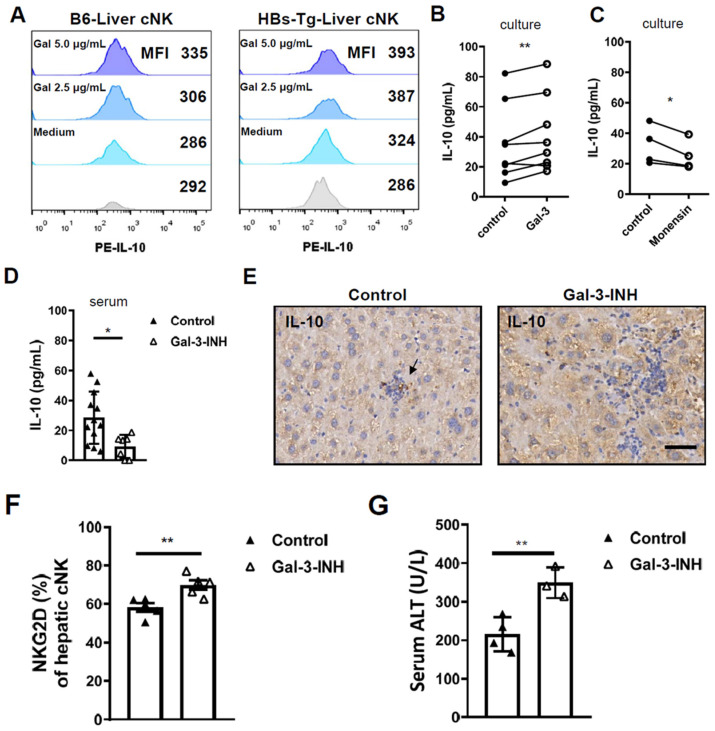
▲ Galectin-3 induced IL-10 production in hepatic cNK cells and prevented hepatocyte damage in HBs-Tg mice. MNCs were isolated from livers of 4- to 5-month-old HBs-Tg mice and control WT B6 mice. (A) IL-10 expression in hepatic cNK cells stimulated by rmGal-3. Liver MNCs (5 × 10⁵) were stimulated by recombinant mouse Gal-3 (2.5 μg/mL or 5 μg/mL) in 200 μL of 10% FBS 1640 medium for 48 h. Cultured MNCs were harvested and analyzed by flow cytometry. cNK cells (CD3⁻NK1.1⁺CD49b⁺CD49a⁻) were gated to analyze IL-10 expression. Histograms and MFI are shown. (B) Levels of IL-10 protein in the culture supernatant. Liver MNCs (5 × 10⁵) were stimulated by recombinant mouse Gal-3 (50 ng/mL) in 400 μL of 10% FBS 1640 medium for 48 h. Culture supernatant was collected and detected by IL-10 CBA flex set. (C) Monensin (2.5 μg/mL) was used to block secretion of IL-10 by cultured NK cells in vitro. Paired Student's t test was used. * p < 0.05, ** p < 0.01. TD139 (Gal-3-INH) (15 μg/g body weight, once every 24 h for three times) was used. (D) Serum levels of IL-10 in HBs-Tg mice after TD139 (Gal-3-INH) treatment detected by ELISA. (E) IL-10 expression in liver tissues detected by IHC. Dark-brown dots represent positive staining. Bar = 50 μm. (F) Hepatic cNK cells (CD3⁻NK1.1⁺CD49b⁺CD49a⁻) were gated and analyzed for expression of NKG2D. (G) Serum levels of ALT after TD139 (Gal-3-INH) treatment. Data are shown as mean ± SEM. Unpaired Student's t test was used. * p < 0.05, ** p < 0.01.
TD139 (T5121) was applied in vivo as a Galectin-3 inhibitor at 15 μg/g body weight, administered once every 24 hours for three doses. In the HBs-Tg mouse model, TD139 produced measurable immunological and hepatic effects. Following TD139 administration, serum interleukin-10 (IL-10) levels were significantly reduced in HBs-Tg mice, demonstrating that IL-10 was sensitive to Galectin-3 inhibition under these conditions. Within the liver compartment, analysis of conventional natural killer (cNK) cells showed that TD139-treated HBs-Tg mice exhibited higher expression of the activating receptor NKG2D on cNK cells, indicating an enhanced activation-associated phenotype linked to Galectin-3 blockade. Systemically, TD139 was associated with elevated serum alanine aminotransferase (ALT) levels in HBs-Tg mice, reflecting increased hepatocyte injury under Galectin-3 inhibition. Collectively, these in vivo findings show that TD139, used to inhibit Galectin-3, decreased circulating IL-10, increased NKG2D expression on hepatic cNK cells, and raised serum ALT in HBs-Tg mice under the dosing regimen described.[4]
■ 3. Conclusion
TD139 functions by competitively binding to the carbohydrate recognition domain of galectin-3 (Gal-3), thereby blocking its interaction with β-galactoside-containing glycoconjugates on cell surface receptors and extracellular matrix components. This inhibition disrupts Gal-3–mediated receptor clustering and signaling lattice formation, attenuating profibrotic signaling pathways such as TGF-β/SMAD and modulating macrophage phenotypes to influence extracellular matrix remodeling. Key studies demonstrate TD139’s efficacy in reducing lysosomal destabilization and inflammasome activation in macrophages, mitigating brain tissue damage and cognitive deficits in ischemic stroke models, and modulating immune responses in hepatitis B virus transgenic mice by decreasing systemic IL-10, enhancing NK cell activation, and causing hepatocyte injury. Future research directions include expanding TD139’s application in fibrotic diseases beyond the lung, exploring its immunomodulatory potential in chronic inflammatory and viral diseases, and refining inhaled delivery methods to optimize local and systemic therapeutic effects.
■ 4. Q&A
Q1: What is the mechanism of action of TD139 in inhibiting Galectin-3?
A1: TD139 competitively binds to the carbohydrate recognition domain of Galectin-3, blocking its interaction with β-galactoside-containing glycoconjugates on cell surface receptors and extracellular matrix, thereby disrupting Gal-3–mediated receptor clustering and signaling lattice formation.
Q2: What effects has TD139 shown in ischemic brain injury models?
A2: TD139 reduced lysosomal destabilization and inflammasome activation in macrophages, mitigated brain tissue loss and edema, improved motor and cognitive functions, and lowered inflammatory cytokine expression after transient middle cerebral artery occlusion in mice.
Q3: How does TD139 affect immune responses in hepatitis B virus transgenic mice?
A3: TD139 decreased serum IL-10 levels, increased activating receptor NKG2D expression on hepatic conventional natural killer cells, and caused elevated serum ALT levels indicative of hepatocyte injury under Galectin-3 inhibition.
■ 5. Reference
[1] Liu FT, Rabinovich GA. Galectins as modulators of immunity. Nat Rev Immunol. 2005;5(1):29-41.
[2] MacKinnon AC, Gibbons MA, Farnworth SL, Leffler H, Nilsson UJ, Delaine T, et al. Regulation of transforming growth factor-β1-driven lung fibrosis by galectin-3. Am J Respir Crit Care Med. 2012;185(5):537-546.
[3] Wang M, Huang Z, Du Z, Shan J, Ye Q, Lu L, et al.. Galectin-3 mediates lysosome-related inflammation within monocyte-derived macrophages in a mouse model of ischemic brain injury. Journal of Clinical Investigation. 2026;136(8):.
[4] Chen Y, Zhang W, Cheng M, Hao X, Wei H, Sun R, et al.. Galectin-3-ITGB1 Signaling Mediates Interleukin 10 Production of Hepatic Conventional Natural Killer Cells in Hepatitis B Virus Transgenic Mice and Correlates with Hepatocellular Carcinoma Progression in Patients. Viruses. 2024;16(5):737.
https://www.targetmol.com/
TargetMol Chemicals Inc.
Post Comment